

Cambridge researchers used lab-grown human mind and spinal twine tissues to uncover a hidden mechanism that blocks nerve restore. By reversing that organic brake, they restored the flexibility of broken nerve fibers to regrow.
Scientists on the College of Cambridge have created miniature mind and spinal twine circuits within the lab that mimic the neural pathways accountable for motion. Utilizing this superior mannequin, they found that injury to those connections, lengthy thought-about everlasting, may very well be reversible.
Because the human physique develops from embryo to fetus and ultimately into infancy, nerve cells known as neurons kind networks that permit alerts to journey between the mind and spinal twine. A vital a part of these cells is the axon, an extended nerve fiber that carries info to different neurons and helps set off muscle motion.
Nonetheless, sooner or later throughout improvement, neurons within the central nervous system lose a lot of their potential to develop new axons. In consequence, injury to the mind or spinal twine usually turns into everlasting, resulting in extreme disabilities corresponding to paralysis or lack of hand operate. This restricted regenerative capability is a serious problem in traumatic spinal twine accidents and neurological issues together with motor neurone illness and a number of sclerosis.
Constructing a Mini Human Mind-Spinal Twine System
In 2021, Dr. András Lakatos and colleagues on the College of Cambridge developed tiny brain-like constructions often known as organoids utilizing human patient-derived stem cells. These stem cells, which might turn into many various cell varieties, have been guided to kind pea-sized, three-dimensional fashions resembling elements of the human cerebral cortex.
The researchers used these early organoids to determine molecular abnormalities concerned in motor neurone illness and discover potential methods to forestall them.
Now, in a research printed in Cell Reports, the team has expanded on that work by creating a miniature version of the interconnected human brain and spinal cord.
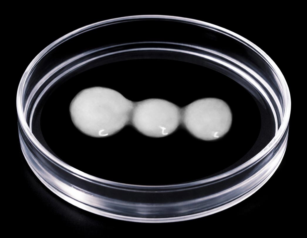
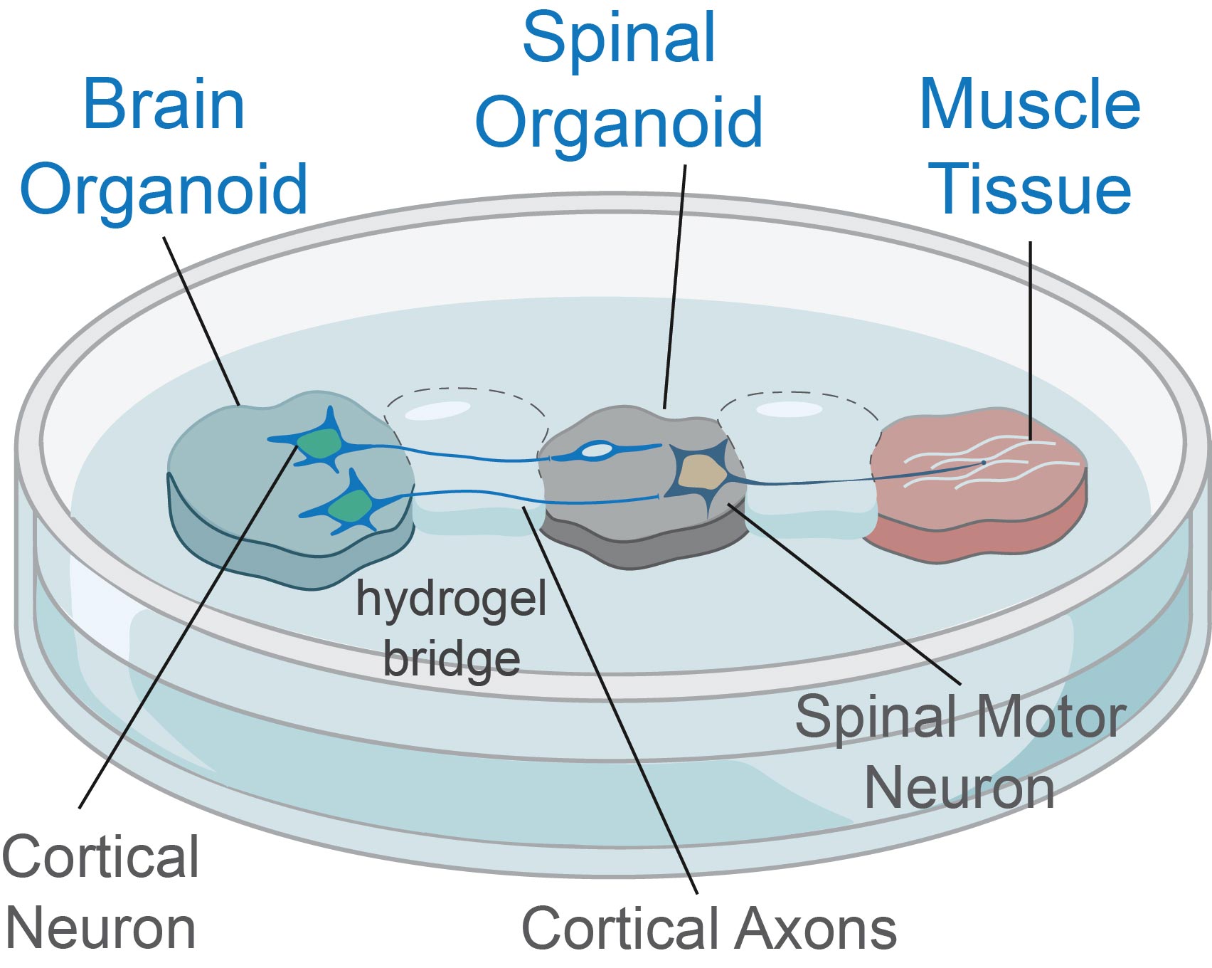
Because the brain and spinal cord are separate structures connected by axons in the body, the researchers grew brain and spinal cord organoids independently. They then observed nerve fibers extending from the brain tissue across a gap and connecting with the spinal cord tissue. The resulting neural circuit was functional enough to trigger contractions in tiny clusters of muscle cells.
When Human Neurons Lose Their Ability to Regrow
The team maintained these miniature nervous systems in the laboratory for more than a year. Their experiments revealed that neurons retained the ability to regrow damaged axons until roughly day 150 of development, which corresponds to the middle stage of pregnancy. After that point, regenerative capacity dropped dramatically.
George Gibbons from the Department of Clinical Neurosciences at the University of Cambridge, the study’s first author, said: “Neurons taken from less mature organoids regrew long fibers after injury, but those from more mature organoids showed a sharp drop in their ability to regrow. In other words, poor regeneration is built into human neurons as they mature in the central nervous system.”
To understand why this happens, the researchers analyzed gene activity in neurons that form connections between the brain and spinal cord. They identified a network of genes that acts like a biological switch, gradually limiting axon growth as neurons mature and establish connections (synapses).
Remarkably, when the scientists blocked key regulators within this gene network, the neurons regained their ability to grow axons.
Existing Drug Boosts Nerve Regeneration
The researchers then searched a database of drug compounds for substances capable of influencing this newly identified genetic network.
One promising candidate was lynestrenol, a hormone drug already approved for treating certain menstrual disorders and for use as a contraceptive.
When the team applied lynestrenol to damaged neurons, axon regrowth increased significantly.
Although scar tissue and inflammation are also known to interfere with nerve repair after injury, the researchers emphasize that understanding neuron-specific barriers is equally important. Previous evidence suggests that younger neurons can often extend axons even through the hostile environments typically found at injury sites.
Senior author Dr. András Lakatos, who led the project at the Department of Clinical Neurosciences, said: “When the brain and spinal cord are damaged, the nerve fibers that carry movement signals from the brain to the spinal cord rarely grow back. That’s why paralysis is usually permanent. But we didn’t know exactly when the ability of axons to regenerate becomes limited. Our model provides a good indication that this block happens during development, and it can still be reversed after this point.
“Lynestrenol itself may not be the answer to spinal cord repair, but it shows us that, in principle, it should be possible to directly target human neurons and regenerate their axons. Although we still need to show that this strategy will also help to re-establish appropriate connections between the brain and spinal cord cells, this gives us hope that one day we may be able to treat conditions previously thought untreatable.”
Human Organoids Help Bridge a Critical Research Gap
Organoids, often called “mini organs,” are becoming increasingly valuable tools for studying human biology and disease.
While animal models such as mice and rats remain important for research, differences between their nervous systems and those of humans can limit how well findings translate to patients. Human organoids offer a closer representation of human biology, helping scientists investigate diseases and treatments in ways that are difficult to achieve with animal studies alone.
Dr. Lakatos added: “Much of what we know about nerve regeneration comes from rodents, whose neurons behave differently from human neurons. Our sophisticated organoid models help bridge the knowledge gap from animal models to what we see in patients. They are also an important contribution to efforts to reduce the use of animals in research.”
Researchers at the University of Cambridge are already using organoids for a wide range of applications, including repairing damaged livers, studying Crohn’s disease in children, and investigating the earliest stages of pregnancy.
Reference: “A human corticospinal organoid-slice connectoid model informs enhancer strategies for post-injury axon regrowth” by George M. Gibbons, Tanja Fuchsberger, Mai Abdelgawad, Stefano L. Giandomenico, Kornélia Szebényi, Veselina Petrova, Lea M.D. Wenger, Daniel N. Olschewski, Jeremi Chabros, Leila Muresan, Rachael C. Feord, Muhammad Asif, James W. Fawcett, Susanna B. Mierau, Ole Paulsen, Madeline A. Lancaster and András Lakatos, 26 May 2026, Cell Reports.
DOI: 10.1016/j.celrep.2026.117399
The study was funded by the UK Research and Innovation Medical Research Council and Spinal Research.
Never miss a breakthrough: Join the SciTechDaily newsletter.
Follow us on Google and Google News.


