
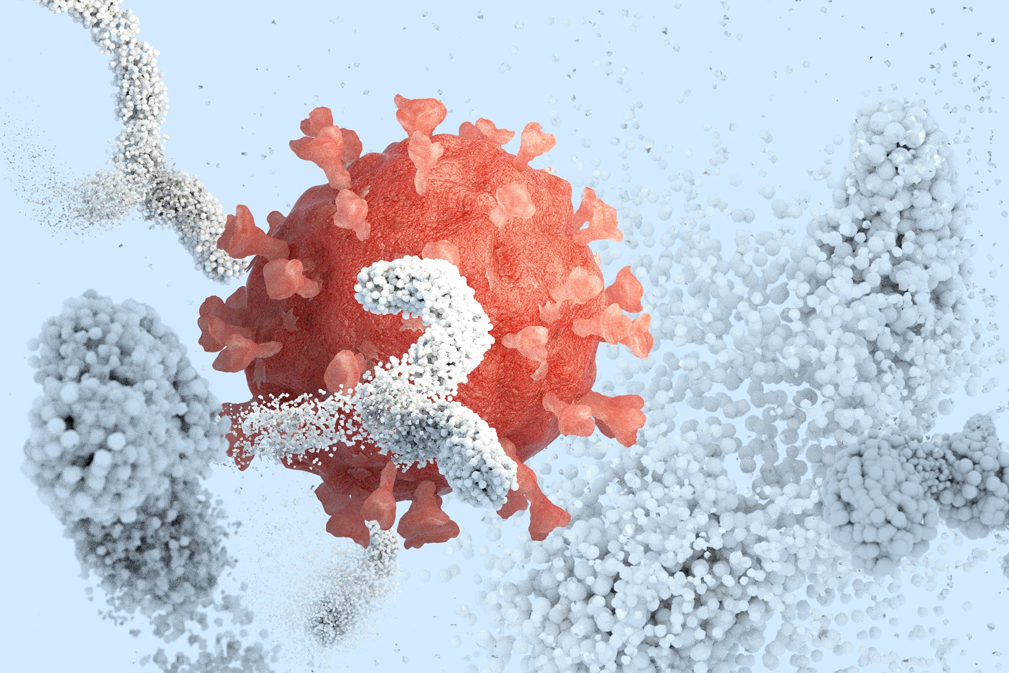
A brand new nanodisc breakthrough lets scientists see viruses extra realistically, revealing hidden clues that would result in higher vaccines.
Viruses are extremely efficient at infecting human cells, largely due to specialised proteins that cowl their outer surfaces. These proteins are additionally a key focus for vaccine design. To review them, scientists typically create lab-made variations to see how the immune system would possibly reply. Nevertheless, these simplified variations normally omit essential sections embedded within the virus’ membrane. Without those pieces, the proteins do not fully behave the way they do in real viruses, making it harder to understand how antibodies recognize and disable them.
Researchers at Scripps Research, working with IAVI and other collaborators, have now developed a new platform that allows these viral proteins to be studied in a form that closely resembles their natural state. The method uses nanodisc technology, where the proteins are placed into tiny particles made of lipids. This creates a membrane-like environment that better preserves their structure and function. As a result, scientists can gain clearer insights into how viral proteins and antibodies interact.
Nanodisc Technology Improves Vaccine Research
The new platform, described in Nature Communications, was tested using proteins from HIV and Ebola. These viruses have been particularly difficult targets for vaccines because their surface proteins are not easily recognized by the immune system. The researchers say the same approach could also be used to study other viruses with similar membrane-bound proteins, including influenza and SARS-CoV-2.
“For many years, we’ve had to rely on versions of viral proteins that are missing important pieces,” says co-senior author William Schief, a professor at Scripps Research and executive director of vaccine design at IAVI’s Neutralizing Antibody Center. “Our platform lets us study these proteins in a setting that better reflects their natural environment, which is critical if we want to understand how protective antibodies recognize a virus.”
Why Membrane Context Matters for Antibodies
In actual viruses, surface proteins are anchored within a lipid membrane and arranged in specific shapes. In contrast, many lab studies remove the membrane-anchoring portion to simplify production and analysis. While this makes experiments easier, it can hide important details. This is especially true for antibodies that target areas close to the base of the protein, near the membrane.
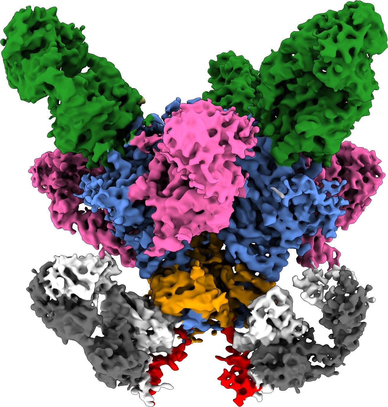
To overcome this limitation, the team incorporated vaccine candidate proteins into nanodiscs, which are small, stable sections of membrane that hold the proteins in place. These lipid discs mimic the outer layer of a virus and help maintain how antibodies would naturally recognize these proteins. The system also supports a wide range of standard techniques used in vaccine development, such as measuring antibody binding, isolating immune cells, and performing high-resolution imaging.
“Putting all of these components together into a single, reliable system was the key,” says first author Kimmo Rantalainen, a senior scientist in Schief’s lab. “The individual pieces already existed, but making them work together in a way that’s reproducible and scalable opens up new possibilities for how vaccines are analyzed and designed.”
New Insights From HIV Antibody Interactions
To test the platform in detail, the researchers focused on HIV. They examined a conserved region of the virus’ surface protein located near the membrane. This area is targeted by a group of antibodies that can block a wide range of HIV variants. These antibodies recognize parts of the virus that stay relatively unchanged even as the virus mutates, making them especially valuable for vaccine design.
Using the nanodisc system, the team captured detailed structural images showing how these antibodies interact with the viral protein in a realistic membrane setting. These images revealed features that cannot be seen when the protein is studied alone. The findings also suggest how certain antibodies neutralize viruses by disrupting the structures they rely on to infect cells. This information could help guide future vaccines to trigger similar immune responses.
“The structure gave us a level of detail we simply couldn’t access before,” notes Rantalainen. “It showed us new interactions at the membrane interface and suggested why those matter for antibody function.”
Expanding Beyond HIV to Other Viruses
The researchers also demonstrated that the platform works with Ebola proteins. In these tests, antibodies were able to recognize and bind to the proteins within the same membrane-like environment, confirming that the approach is not limited to a single virus.
A Faster Way To Study Immune Responses
Beyond analyzing structures, the platform can also be used to study how the immune system responds to vaccine candidates. The nanodiscs can act as molecular “bait,” allowing scientists to isolate immune cells that recognize viral proteins. This provides a clearer picture of how the body reacts to different vaccine designs. The system is also faster and more efficient. Processes that once took a month or longer can now be completed in about a week, making it easier to compare multiple vaccine candidates.
A Tool To Advance Next Generation Vaccines
Although this platform is not itself a vaccine, it offers a powerful tool to improve how vaccines are developed. It may be especially useful for viruses that have proven difficult to target using traditional methods.
“This gives the field a more realistic, accurate way to test ideas early on,” emphasizes Schief. “By improving how we study viral proteins and antibody responses, we hope this platform will help advance next-generation vaccines against some of the world’s most challenging viruses.”
Reference: “Virus glycoprotein nanodisc platform for vaccine analytics” by Kimmo Rantalainen, Alessia Liguori, Gabriel Ozorowski, Claudia Flynn, Jon M. Steichen, Olivia M. Swanson, Patrick J. Madden, Sabyasachi Baboo, Swastik Phulera, Anant Gharpure, Danny Lu, Oleksandr Kalyuzhniy, Patrick Skog, Sierra Terada, Monolina Shil, Jolene K. Diedrich, Erik Georgeson, Ryan Tingle, Saman Eskandarzadeh, Wen-Hsin Lee, Nushin Alavi, Diana Goodwin, Michael Kubitz, Sonya Amirzehni, Sunny Himansu, Devin Sok, Jeong Hyun Lee, John R. Yates III, James C. Paulson, Shane Crotty, Torben Schiffner, Andrew B. Ward and William R. Schief, 10 February 2026, Nature Communications.
DOI: 10.1038/s41467-026-68985-1
In addition to Schief and Rantalainen, authors of the study include Alessia Liguori, Gabriel Ozorowski, Claudia Flynn, Jon M. Steichen, Olivia M. Swanson, Patrick J. Madden, Sabyasachi Baboo, Swastik Phulera, Anant Gharpure, Danny Lu, Oleksandr Kalyuzhniy, Patrick Skog, Sierra Terada, Monolina Shil, Jolene K. Diedrich, Erik Georgeson, Ryan Tingle, Saman Eskandarzadeh, Wen-Hsin Lee, Nushin Alavi, Diana Goodwin, Michael Kubitz, Sonya Amirzehni, Devin Sok, Jeong Hyun Lee, John R. Yates III, James C. Paulson, Shane Crotty, Torben Schiffner and Andrew B. Ward of Scripps Research; and Sunny Himansu of Moderna Inc.
This work was supported by funding from the National Institute of Allergy and Infectious Diseases of the National Institutes of Health (grants UM1 AI144462, R01 AI147826, R56 AI192143 and 5F31AI179426-02); the Bill and Melinda Gates Foundation Collaboration for AIDS Vaccine Discovery (grants INV-007522, INV-008813 and INV-002916); the IAVI Neutralizing Antibody Center (INV-034657 and INV-064772); and the Alexander von Humboldt Foundation.
Never miss a breakthrough: Join the SciTechDaily newsletter.
Follow us on Google and Google News.


